
Radiology Ordering Guide: A Comprehensive Overview (Updated 12/31/2025)
This guide‚ updated December 31st‚ 2025‚ assists providers in ordering effective imaging‚ enhancing patient care‚ and utilizing radiology resources efficiently.
Radiology ordering is a critical component of modern healthcare‚ directly impacting diagnostic accuracy and patient management. This guide serves as a comprehensive resource for referring providers‚ aiming to streamline the ordering process and ensure appropriate test selection. Effective radiology utilization requires a clear understanding of clinical indications‚ imaging modalities‚ and established guidelines.
Developed by radiologists at Radia‚ these guidelines promote quality of care and efficient resource allocation. Proper ordering minimizes unnecessary radiation exposure‚ reduces costs‚ and ultimately improves patient outcomes. This document will detail best practices for various clinical scenarios‚ assisting you in selecting the most effective imaging approach for each individual case.
II. Importance of Appropriate Test Selection

Appropriate test selection in radiology is paramount for delivering optimal patient care. Ordering the correct imaging study‚ based on specific clinical indications‚ avoids unnecessary radiation exposure and minimizes diagnostic delays. Utilizing the least invasive modality first‚ as guidelines suggest‚ is a core principle.

Inappropriate ordering leads to increased healthcare costs‚ potential harm to patients‚ and inefficient use of radiology resources. This guide emphasizes the importance of carefully considering the clinical question being asked and selecting the imaging technique most likely to provide a definitive answer. Collaboration with radiologists is encouraged to ensure the most appropriate study is chosen.
III. Understanding Radiology Guidelines
Radiology guidelines are crucial for standardizing care and optimizing imaging practices. These protocols‚ developed by radiologists at Radia‚ aim to enhance the quality of care provided to patients. Adherence to these guidelines ensures efficient utilization of radiology resources‚ reducing unnecessary examinations and associated costs.
Understanding these recommendations allows providers to select the most effective imaging modality for each clinical scenario. The guidelines are regularly updated to reflect advancements in technology and best practices‚ promoting evidence-based ordering. Consistent application of these standards contributes to improved diagnostic accuracy and patient outcomes.
A. Quality of Care Enhancement
Radiology guidelines directly contribute to enhanced quality of care by promoting appropriate test selection. Utilizing these protocols ensures patients receive the most clinically relevant imaging‚ leading to more accurate diagnoses and tailored treatment plans. By following established guidelines‚ providers minimize patient exposure to radiation and reduce the likelihood of incidental findings requiring further investigation.
This focused approach improves diagnostic confidence and facilitates timely interventions‚ ultimately improving patient outcomes. Consistent adherence to these standards fosters a culture of quality within healthcare facilities‚ benefiting both patients and providers.
B. Efficient Use of Radiology Resources
Radiology resources are valuable and finite; therefore‚ efficient utilization is paramount. Following established guidelines minimizes unnecessary examinations‚ reducing wait times for all patients and optimizing the scheduling of imaging procedures. This careful approach also lowers healthcare costs associated with imaging services‚ benefiting both patients and the healthcare system.
By selecting the most appropriate modality for each clinical indication‚ we avoid redundant testing and ensure that radiologists can focus on cases requiring their expertise. This streamlined process improves workflow and maximizes the impact of available resources.

IV. The Role of the Radiologist
Radiologists play a crucial role beyond image acquisition and interpretation; they are vital partners in patient care. Their expertise ensures accurate diagnoses and guides treatment decisions. Findings and recommendations are meticulously communicated to referring providers‚ offering comprehensive insights into the patient’s condition.
Effective communication between the radiologist and the referring provider is essential for optimal patient outcomes. This includes clear articulation of imaging results‚ potential diagnoses‚ and suggested follow-up actions‚ fostering a collaborative approach to healthcare.
A. Findings and Recommendations
Radiologist findings are detailed reports outlining observations from the imaging study‚ encompassing both normal anatomy and any identified abnormalities. These reports aren’t simply descriptive; they translate complex imaging data into clinically relevant information for the referring physician.
Recommendations build upon these findings‚ suggesting appropriate next steps – whether it’s further imaging‚ correlation with clinical data‚ or referral to a specialist. These recommendations are crucial for guiding patient management and ensuring timely‚ effective care. For example‚ an order for diagnostic mammogram with breast ultrasound may be suggested.

B. Communication with Referring Providers
Effective communication between radiologists and referring providers is paramount for optimal patient care. Radiologists don’t operate in isolation; they are integral members of the healthcare team. Prompt and clear reporting of findings‚ coupled with readily available consultation‚ ensures informed decision-making.
This includes not only delivering the written report but also being accessible to discuss complex cases‚ clarify recommendations‚ and answer questions. Open dialogue fosters a collaborative environment‚ leading to more accurate diagnoses and tailored treatment plans. This collaborative approach ultimately benefits the patient.
V. General Ordering Principles
Adhering to general ordering principles is crucial for responsible radiology utilization. Begin with the least invasive imaging modality possible‚ considering the clinical question and potential radiation exposure. Prioritize modalities like ultrasound or radiography before proceeding to CT or MRI when appropriate.
Specificity in clinical indication is also vital. A detailed and precise order ensures the radiologist can tailor the protocol to the specific clinical concern‚ maximizing diagnostic yield. Avoid vague requests; instead‚ clearly articulate the suspected pathology or clinical question driving the imaging request.
A. Starting with Least Invasive Modalities
Prioritizing less invasive techniques minimizes patient risk and exposure to ionizing radiation. Begin with modalities like radiography (X-ray) or ultrasound‚ which lack ionizing radiation‚ whenever clinically feasible. These options often provide valuable diagnostic information for many conditions.
If further evaluation is needed‚ consider CT scans before MRI‚ balancing diagnostic yield with radiation dose. Always justify the need for higher-risk modalities based on the clinical indication. This approach aligns with the ALARA (As Low As Reasonably Achievable) principle‚ safeguarding patient well-being.
B. Clinical Indication Specificity
Precise clinical details are crucial for appropriate image selection and interpretation; Vague requests like “rule out pathology” are insufficient; specify the suspected diagnosis or clinical question. Detailed information guides radiologists toward the most relevant imaging protocol‚ reducing unnecessary scans and improving diagnostic accuracy.
Clearly articulate the patient’s symptoms‚ relevant history‚ and prior imaging results. This context enables radiologists to tailor the exam to the specific clinical scenario‚ optimizing image quality and minimizing potential for misinterpretation. Accurate ordering directly impacts patient care.
VI. Medicare Ordering Requirements
Medicare mandates explicit‚ written‚ and signed provider orders for all radiology examinations. Verbal orders are not acceptable for reimbursement. The order must clearly detail the specific imaging procedure requested‚ including laterality when applicable‚ and the corresponding clinical indication justifying the exam.
Ensure orders adhere to Medicare’s documentation guidelines to avoid claim denials. This guide assists in accurate exam authorization. Proper documentation supports medical necessity and ensures appropriate utilization of radiology services‚ aligning with Medicare’s quality and efficiency standards.
VII. Breast Imaging Guidelines
Breast imaging protocols require careful consideration. For diagnostic evaluations‚ consider ordering a diagnostic mammogram coupled with a breast ultrasound for comprehensive assessment. If a patient declines the 3D imaging protocol‚ a diagnostic 2D mammogram is appropriate for five years‚ followed by a return to screening in the sixth year.
Always assess if the patient has current breast concerns or changes. Following these guidelines ensures appropriate imaging selection and adherence to best practices for early detection and accurate diagnosis‚ ultimately improving patient outcomes.

A. Diagnostic Mammography & Ultrasound
Diagnostic mammography is crucial when investigating specific breast concerns‚ such as a palpable lump or nipple discharge. Often‚ it’s optimally paired with a breast ultrasound to differentiate between solid and cystic masses‚ providing complementary information. This combined approach enhances diagnostic accuracy.
Ultrasound is particularly valuable for evaluating dense breast tissue where mammography’s sensitivity may be reduced. Ordering both modalities ensures a thorough evaluation‚ leading to earlier and more precise diagnoses‚ ultimately guiding appropriate patient management and treatment plans.
B. 3D Imaging Protocols & Alternatives
3D mammography‚ or tomosynthesis‚ offers a more comprehensive breast evaluation by creating multiple images from different angles‚ reducing tissue overlap and improving cancer detection rates. However‚ patient preference or facility limitations may necessitate alternatives.
If a patient declines 3D imaging‚ a diagnostic 2D mammography protocol is recommended for five years‚ followed by a return to screening in the sixth year. Careful consideration of individual risk factors and clinical presentation is vital when selecting the most appropriate imaging modality for each patient.
C. Screening Mammography Follow-up
Appropriate follow-up after screening mammography is crucial for ensuring optimal patient outcomes. Recommendations vary based on findings and individual risk factors. Standard follow-up may include short-interval surveillance‚ diagnostic mammography with ultrasound‚ or biopsy if suspicious abnormalities are detected.
Providers should clearly communicate follow-up instructions to patients and document all recommendations in the patient’s medical record. Consistent adherence to established guidelines and prompt investigation of any concerning findings are essential components of effective breast cancer screening programs.
VIII. Musculoskeletal Radiology Ordering
Musculoskeletal radiology encompasses imaging of bones‚ joints‚ muscles‚ and soft tissues. When ordering‚ specify the clinical indication precisely. For Temporomandibular Joint (TMJ) pain‚ MRI without contrast utilizing a dedicated TMJ protocol is often the initial modality of choice‚ particularly in non-traumatic cases.
Clearly document the patient’s symptoms and relevant history on the order. Consider alternative imaging modalities if MRI is contraindicated. Accurate clinical information guides the radiologist in selecting the most appropriate imaging technique and interpreting the findings effectively‚ leading to a precise diagnosis.
A. MRI for Temporomandibular Joint (TMJ) Pain
MRI without contrast‚ utilizing a specific TMJ protocol‚ is the preferred initial imaging modality for non-traumatic temporomandibular joint (TMJ) pain. This protocol focuses on detailed visualization of the joint structures‚ including the disc‚ cartilage‚ and surrounding muscles.
When ordering‚ clearly indicate “MRI without contrast (TMJ protocol)” to ensure the correct sequence is performed. Document the duration and nature of the patient’s pain‚ any clicking or locking sensations‚ and limitations in jaw movement. This detailed clinical information assists the radiologist in accurate interpretation and diagnosis.
IX. Neck Imaging Guidelines
Neck imaging guidelines focus on appropriate evaluation of palpable masses‚ excluding thyroid abnormalities. For a non-thyroid mass‚ a comprehensive clinical history is crucial when ordering diagnostic exams. Document the mass’s location‚ size‚ consistency‚ and any associated symptoms like pain or dysphagia.
Initial evaluation typically involves ultrasound‚ providing excellent soft tissue detail. If ultrasound findings are inconclusive or concerning‚ a CT scan with contrast or MRI may be necessary for further characterization. Always specify the patient symptoms when submitting the order for optimal interpretation.
A. Mass (Non-Thyroid) Evaluation
Evaluating a non-thyroid neck mass requires a systematic approach. Begin with a detailed patient history‚ noting duration‚ growth rate‚ and associated symptoms. Ultrasound is generally the first-line imaging modality‚ effectively differentiating cystic from solid lesions.
For solid masses‚ consider CT with contrast or MRI to assess size‚ location‚ and involvement of adjacent structures. Clearly document patient symptoms – such as pain or dysphagia – on the order. This guides radiologist interpretation and ensures appropriate follow-up recommendations. Accurate clinical information is paramount for precise diagnosis.
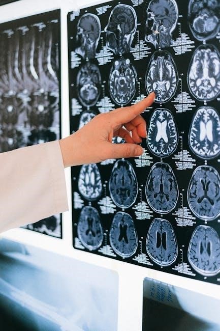
X. Neurological Radiology Ordering
Neurological imaging requires precise order details. For suspected stroke‚ immediate non-contrast CT is crucial to differentiate ischemic from hemorrhagic events. MRI with and without contrast is preferred for evaluating soft tissue abnormalities‚ spinal cord lesions‚ and demyelinating diseases.
Clearly indicate clinical suspicion – headache‚ weakness‚ seizures – on the order. Specify relevant history‚ such as prior stroke or aneurysm. Consider MR angiography (MRA) or CT angiography (CTA) for vascular assessment. Radiologist consultation regarding protocol selection is encouraged for complex cases‚ optimizing diagnostic yield.
XI. Abdominal and Pelvic Imaging Guidelines
Abdominal and pelvic imaging selection depends heavily on clinical presentation. For acute abdominal pain‚ CT with IV contrast is often the initial modality‚ evaluating for appendicitis‚ diverticulitis‚ and bowel obstruction. Ultrasound is valuable for gallbladder and biliary tract assessment‚ particularly in pregnant patients.
MRI provides superior soft tissue detail for evaluating liver lesions‚ pancreatic masses‚ and pelvic pathology. Specify relevant history – prior surgeries‚ cancer – on the order. Consider oral and IV contrast optimization. Radiologist consultation aids in protocol tailoring for specific clinical questions.
XII. Chest Radiology Ordering
Chest radiography remains the initial imaging modality for evaluating pulmonary symptoms like cough‚ shortness of breath‚ and chest pain. For suspected pneumonia‚ a PA and lateral view are standard. CT chest with and without contrast is indicated for more complex cases – pulmonary embolism‚ lung cancer staging‚ or interstitial lung disease;
Specify clinical suspicion on the order; for example‚ “rule out PE” or “evaluate for lung nodule.” Consider prior imaging for comparison. Be mindful of radiation exposure‚ especially with repeated CT scans. Radiologist consultation can refine protocol selection.
XIII. Pediatric Radiology Considerations
Pediatric imaging requires careful consideration of radiation dose minimization. Utilize the ALARA (As Low As Reasonably Achievable) principle. Protocols should be age-appropriate and adjusted for body habitus. Shielding is crucial‚ particularly for gonadal and thyroid regions.
Clinical history is paramount; include relevant details like growth parameters and developmental milestones on the order. Sedation or general anesthesia may be necessary for younger or uncooperative patients. Prioritize ultrasound and MRI when feasible to avoid ionizing radiation. Consult with a pediatric radiologist when possible.

XIV. Contrast Agent Considerations
Prior to contrast administration‚ a thorough allergy history is essential. Document any previous reactions to contrast agents‚ medications‚ or food allergies. Assess renal function via estimated glomerular filtration rate (eGFR) or creatinine levels‚ especially in patients with diabetes or pre-existing kidney disease.
Hydration protocols may be necessary to minimize the risk of contrast-induced nephropathy. Consider alternative imaging modalities if renal function is significantly impaired. Always inform patients about potential side effects and obtain informed consent before administering contrast.
A. Allergy History
A comprehensive allergy history is paramount before administering contrast agents. Specifically document any prior reactions to iodinated contrast‚ medications‚ or food allergies‚ noting the type of reaction experienced. Even seemingly unrelated allergies should be recorded‚ as cross-reactivity can occur.
Patients reporting a history of contrast reaction require careful evaluation and potentially pre-medication with corticosteroids and antihistamines. Clearly document the allergy status on the order to alert the radiologist and technologist.
B. Renal Function Assessment
Prior to contrast-enhanced studies‚ assessing renal function is crucial to mitigate the risk of contrast-induced nephropathy (CIN). Obtain a recent serum creatinine level and calculate the estimated glomerular filtration rate (eGFR). Document these values clearly on the radiology order.
Patients with impaired renal function (low eGFR) may require hydration protocols or alternative imaging modalities to avoid CIN. Radiologists may adjust contrast dosage based on renal function. Failure to assess renal function can lead to adverse patient outcomes;
XV. Patient Preparation Instructions
Clear communication of preparation instructions is vital for optimal image quality and accurate diagnoses. Specific instructions vary based on the exam; therefore‚ providers must inform patients accordingly. For example‚ bowel preparation is often required for abdominal and pelvic CT scans.
Patients should receive written and verbal instructions‚ including dietary restrictions‚ medication adjustments‚ and arrival times. Ensure patients understand the importance of following these guidelines. Failure to comply can necessitate rescheduling‚ delaying diagnosis and treatment. Detailed instructions improve exam success.
XVI. Documentation Requirements for Orders

Complete and accurate documentation is paramount for all radiology orders. Medicare guidelines mandate explicit‚ written‚ and signed provider orders‚ detailing the clinical indication for the requested exam. Vague or incomplete orders may lead to denials or delays in authorization.
Orders must include relevant patient history‚ physical exam findings‚ and prior imaging reports. Justification for the chosen modality should be clearly stated‚ demonstrating medical necessity. Proper documentation ensures appropriate utilization of resources and supports accurate billing practices‚ safeguarding both patient care and financial integrity.

XVII. Order Authorization Processes
Prior authorization may be required for certain radiology examinations‚ dictated by payer policies and medical necessity criteria. This guide assists in accurately ordering and authorizing exams. Providers are responsible for verifying authorization requirements before scheduling the procedure.
Authorization requests typically necessitate detailed clinical information‚ supporting documentation‚ and a clear justification for the requested imaging. Electronic submission is often preferred for efficiency. Failure to obtain required authorization can result in claim denials. Prompt and accurate authorization streamlines the process‚ ensuring timely patient access to necessary care.
XVIII. Common Ordering Errors to Avoid
Avoid vague clinical indications; specificity is crucial for appropriate exam selection and authorization. Incorrect CPT codes or missing information frequently cause delays and denials. Ensure patient demographics and insurance details are accurate on each order.
Duplication of prior imaging without justification should be avoided‚ as it exposes patients to unnecessary radiation. Failure to consider alternative modalities‚ starting with the least invasive‚ is a common error. Regularly review and adhere to established radiology guidelines to minimize errors and optimize patient care.
XIX. Utilizing Decision Support Tools
Decision support tools integrated within Electronic Health Records (EHRs) can significantly improve radiology ordering. These tools often provide guidelines based on clinical indications‚ assisting providers in selecting the most appropriate exam.
Automated alerts can flag potentially inappropriate orders‚ such as those lacking sufficient clinical detail or violating established protocols. Utilizing these resources promotes adherence to best practices‚ reduces unnecessary imaging‚ and streamlines the order authorization process‚ ultimately enhancing both quality of care and resource utilization.
XX. Staying Updated on Radiology Guidelines
Radiology guidelines are continuously evolving with advancements in technology and clinical research. Providers must commit to ongoing education to ensure ordering practices remain current and aligned with best practices.
Regularly reviewing updates from professional societies‚ such as the American College of Radiology (ACR)‚ and participating in continuing medical education (CME) activities are crucial. Staying informed about changes in Medicare requirements and local hospital protocols is also essential for accurate ordering and appropriate patient care.
XXI. Resources for Ordering Information
Radia provides comprehensive resources to support informed radiology ordering. Our website features detailed scanning guidelines‚ clinical decision support tools‚ and frequently updated protocol information for various imaging modalities.
Referring providers can access these resources 24/7. Additionally‚ our radiology support team is available to answer questions and provide assistance with order authorization processes. For Medicare specific guidance‚ refer to the CMS website. Utilizing these tools ensures accurate ordering and optimal patient outcomes.
XXII. Contacting Radiology Support
Radia’s dedicated radiology support team is readily available to assist referring providers with any ordering questions or concerns. We strive to ensure a seamless and efficient process for all imaging requests.
For immediate assistance‚ please call our support line at 555-123-4567 during business hours (8 AM ⎼ 5 PM‚ Monday-Friday). Alternatively‚ you can email us at radiologysupport@radia.com. We are committed to providing prompt and helpful support to facilitate optimal patient care and accurate order authorization.